|
#2
September 30th, 2016, 09:15 AM
| |||
| |||
| Re: Previous AIIMS PG Question Papers
Well below I have given you the previous AIIMS PG question papers so you can have a look ANATOMY 1. In the advancement of surgery of shoulder joint, one muscle was not given much importance earlier but now came in picture, this forgotten muscle of rotator cuff is a. Subscapularis b. Supraspinatus c. Infraspinatus d. Teres minor Answer: (a) Subscapularis Reference: Explanation:Musculotendinous cuff of shoulder is a fibrous sheath formed by the four flattened tendons which blend with the capsule of shoulder joint and strengthen it. Muscles which form the cuff arise from scapula and are inserted into the lesser and greater tubercles of humerus. They are subscapularis, supaspintaous, infraspinatus and teres minor. There tendons while crossing the shoulder joint become flattened and blend with each other on one end and with capsule of the joint on the other end, before reaching their points of insertion.Cuff gives strength to the capsule of shoulder joint all around except inferiorly. That is why dislocation of humerusoccur most commonly in a down ward insertion. 2. Stuctures at anorectal junction all except a. External sphincter b. Internal sphincter c. Anococcygeal raphe d. Puborectalis Answer (c)Anococcygealraphae Reference: BDC 2nd vol,5/e, pg.414 Explanation: Anorectal ring is a muscular ring present at the anorectal junction. It is formed by the fusion of the puborectalis, uppermost fibers of external sphincter and internal sphincter. Surgical division of anorectal ring results in rectal incontinence. Anococcygeal ligament or anococcygeal raphe is a multilayer musculotendinous structure between anal canal & hip of coccyx. It is attached anteriorly to the superficial part of external anal sphincter i.e. middle part of external anal sphincter which lies below to deep part of external anal sphincter at the anorectal junction present deep part of external anal sphincter. 3. Structure not present at floor of third ventricle- a. Optic stalk b. Third nerve c. Infundibulum d. Mamillary body Answer: (b) Third nerve (Occulo motor N.) Reference: BDC 3rdvol, 5/e, pg. 428 Explanation: Third ventricle is a median cleft b/w two thalami. Embryologically it represents the cavity of diencephalon. Communication :Antero superiorly, on each side it communicates with the lateral ventricle through the interventriclar foramen (or Foramen of Monro). Postero inferiorly it communicates with the fourth ventricle through the Cerebral Aqueduct. Boundaries: Anterior wall – i. Lamina terminalis ii. Anterior commissure iii. Anterior columns of fornix Posterior wall – i. Pineal body ii. Post commissure iii. Cerebral aqueduct Roof - i. Ependymal liningof the under surface of telachoroidea of third ventricle. Floor – i. Optic chiasma ii. Tubercinerium iii. Infndibulum (Pituitary stalk) iv. Mamillary bodies v. Posterior perforated substance vi. Tegmentum of the mid rain Lateral wall – i. Medial surface of thalamus ii. Hypothalamus iii. Hypothalamic sulcus Occulomotor N arises at the medial end of crus cerebri of mid brain.So it is not present at the floor of IIIrd ventricle 4. Which among the following forma a complete cartilaginous ring around tracheobronchial tree- a. Cricoidcartilage b. Epiglottis c. Cuneiformcartilage d. Thyroid cartilage Answer (a) Cricoids cartilage Ref: BDC 3rdvol, 5/e pg. 238 Explanation: Cricoid cartilage is shaped like a ring. It encircles the larynx below the thyroid cartilage. The ring has a narrow anterior part called the arch and a broad posterior part called the lamina. Lamina projects upwards behind the thyroid cartilage and articulates superiorly with the arytenoids cartilages. Larynx contains nine cartilages out of which 3 are paired & 3 are unpaired. Unpaired cartilages i. Thyroid – V shaped ii. Cricoids – Ring shaped iii. Epiglottic – Leaf shaped Paired cartilages i. Arytenoids – Pyramid shaped ii. Corniculate – Small conical shape iii. Cuneiform – Small rod shape 5. Father of neuro-otology was a. William House b. John shea c. Joseph lampert d. Not recalled Answer: (a) William F. HouseInMemorian: Reference: Wiliam F. House, D.D.S. M.D. the “Father of Neurotology” 1923 – 2012 Explanation: Berlinger, Karen Otology and Neurotology 34 (3): 386 – 387 April 2013 Otology &Neurotology is the name of journal. In which this article has been published available on internet. 6. Vaginal endothelium is derived from: a. Endoderm of urogenital sinus b. Mesoderm of urogenital sinus c. Endoderm of genital ridge d. Mesoderm of genital ridge Ans: A, Endoderm of urogenital sinus Ref: BDC 2ndvol, 5/e pg. 397 Exp: As the fused Müllerian or paramesonephric ducts which form the uterovaginal canal open into the definitive urogenital sinus, the endoderm bulges to form the Müllerian tubercle. Uterovaginal canal forms upper third of vagina. Endoderm on either side of Müllerian tubercle proliferates to form two sinovaginal bulbs which fuse to form vaginal plate. The vaginal plate surrounds the caudal end of the uterovaginal canal. Soon there is a canalization of the vaginal plate to form lower 2/3rd of vagina and vaginal fornices. It opens through an endodermal partial septum- the hymen in definitive urogenital sinus. 7. Floor of orbit is not formed by all except a. Ethmoid b. Maxilla c. Zygomatic d. Palatine Ans: A,Ethmoid Ref: [REF TEXTBOOK OF HUMAN OSTEOLOGY INDERBIR SING 2/E P-128-130; BDC 4/E VOL III P. 27] Exp: MEDIAL WALL OF ORBIT IS FORMED BY MAXILLA, SPHENOID, ETHMOID & LACRIMAL BONE Previous AIIMS PG Question Papers     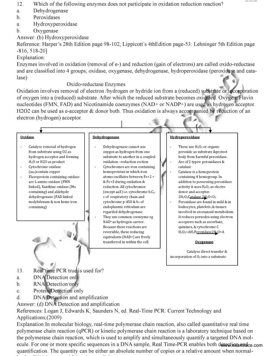 |