|
#2
April 9th, 2017, 11:26 AM
| |||
| |||
| Re: AP PGMET Key
As you Asking for the Solved Question Paper of the AP PGMET Exam Let me tell you that the AP PGMET Exam is Replaced With the NEET PG Exam the 1. In the advancement of surgery of shoulder joint, one muscle was not given much importance earlier but now came in picture, this forgotten muscle of rotator cuff is a. Subscapularis b. Supraspinatus c. Infraspinatus d. Teres minor Answer: (a) Subscapularis Reference: Explanation:Musculotendinous cuff of shoulder is a ibrous sheath formed by the four lattened tendonswhich blend with the capsule of shoulder joint and strengthen it. Muscles which form the cuff arise from scapula and are inserted into the lesser and greater tubercles of humerus. They are subscapularis, supaspintaous, infraspinatus and teres minor. There tendons while crossing the shoulder joint become lattened and blend with each other on one end and with capsule of the joint on the other end, before reaching their points of insertion.Cuff gives strength to the capsule of shoulder joint all around except inferiorly. That is why dislocation of humerusoccur most commonly in a down ward insertion. 2. Stuctures at anorectal junction all except a. External sphincter b. Internal sphincter c. Anococcygeal raphe d. Puborectalis Answer (c)Anococcygealraphae Reference: BDC 2nd vol,5/e, pg.414 Explanation: Anorectal ring is a muscular ring present at the anorectal junction. It is formed by the fusion of the puborectalis, uppermost ibers of external sphincter and internal sphincter. Surgical division ofanorectal ring results in rectal incontinence. Anococcygeal ligament or anococcygeal raphe is a multilayer musculotendinous structure between anal canal & hip of coccyx. It is attached anteriorly to the supericial part of external anal sphincter i.e. middle part of external anal sphincter which lies below to deep part of external anal sphincter at the anorectaljunction present deep part of external anal sphincter. 3. Structure not present at loor of third ventricle- a. Optic stalk b. Third nerve c. Infundibulum d. Mamillary body Answer: (b) Third nerve (Occulo motor N.) Reference: BDC 3rdvol, 5/e, pg. 428 Explanation: Third ventricle is a median cleft b/w two thalami. Embryologically it represents the cavity of diencephalon. Communication :Antero superiorly, on each side it communicates with the lateral ventricle through the interventriclar foramen (or Foramen of Monro). Postero inferiorly it communicates with the fourth ventricle through the Cerebral Aqueduct. Boundaries: Anterior wall – i. Lamina terminalis ii. Anterior commissure iii. Anterior columns of fornix Posterior wall – i. Pineal body ii. Post commissure iii. Cerebral aqueduct Roof - i. Ependymal liningof the under surface of telachoroidea of third ventricle. Floor – i. Optic chiasma ii. Tubercinerium iii. Infndibulum (Pituitary stalk) iv. Mamillary bodies v. Posterior perforated substance vi. Tegmentum of the mid rain Lateral wall – i. Medial surface of thalamus ii. Hypothalamus iii. Hypothalamic sulcus Occulomotor N arises at the medial end of crus cerebri of mid brain.So it is not present at the loorof IIIrd ventricle 4. Which among the following forma a complete cartilaginous ring around tracheobronchial tree- a. Cricoidcartilage b. Epiglottis c. Cuneiformcartilage d. Thyroid cartilage Answer (a) Cricoids cartilage Ref: BDC 3rdvol, 5/e pg. 238 Explanation: Cricoid cartilage is shaped like a ring. It encircles the larynx below the thyroid cartilage. The ring has a narrow anterior part called the arch and a broad posterior part called the lamina. Lamina projects upwards behind the thyroid cartilage and articulates superiorly with the arytenoids cartilages. Larynx contains nine cartilages out of which 3 are paired & 3 are unpaired. Unpaired cartilages i. Thyroid – V shaped ii. Cricoids – Ring shaped iii. Epiglottic – Leaf shaped Paired cartilages i. Arytenoids – Pyramid shaped ii. Corniculate – Small conical shape iii. Cuneiform – Small rod shape 5. Father of neuro-otology was a. William House b. John shea c. Joseph lampert d. Not recalled Answer: (a) William F. HouseInMemorian: AP PGMET Exam Question Paper    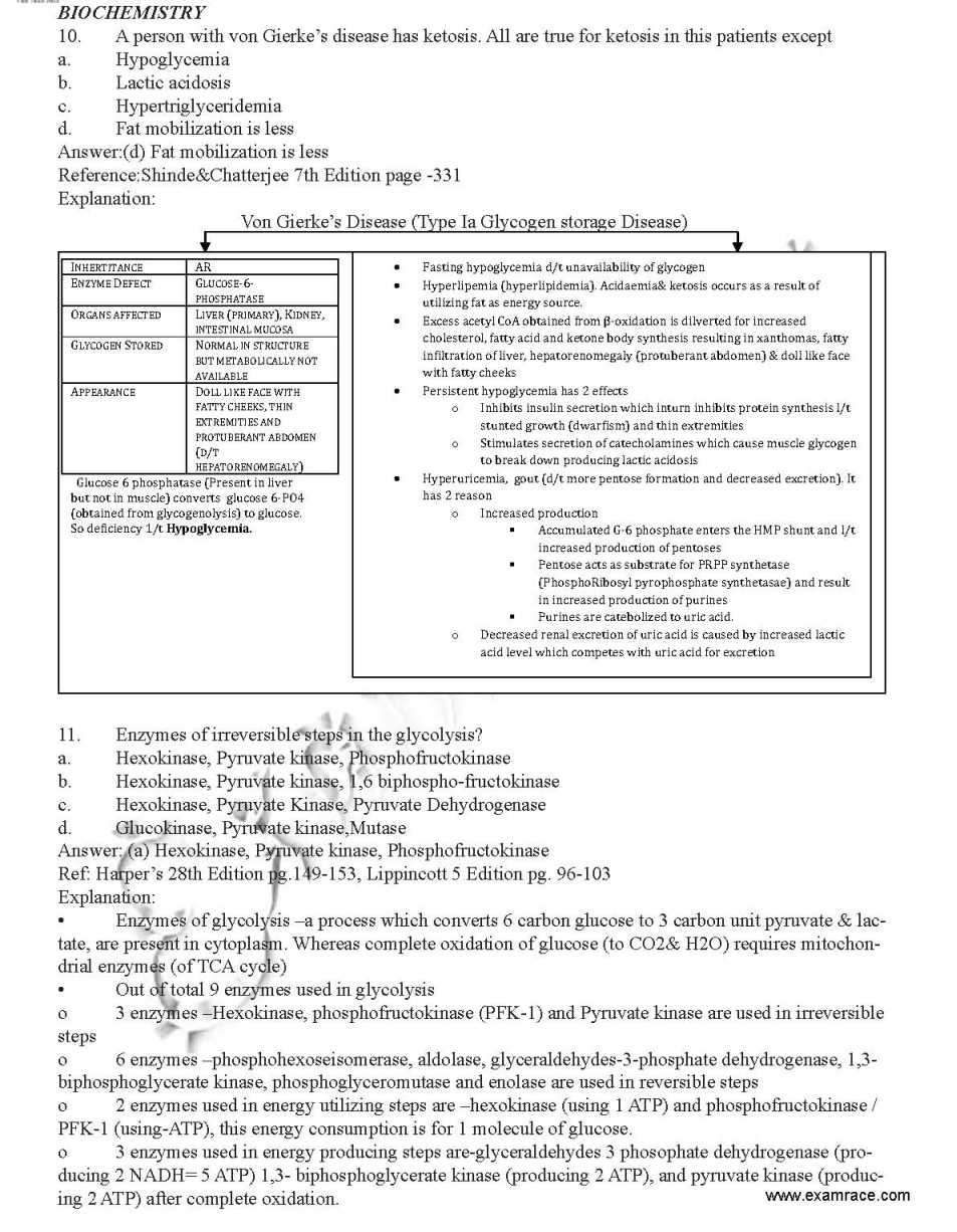 Rest of the Question Paper you May get From the below Attachement that is Free to Download |